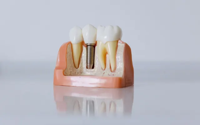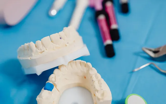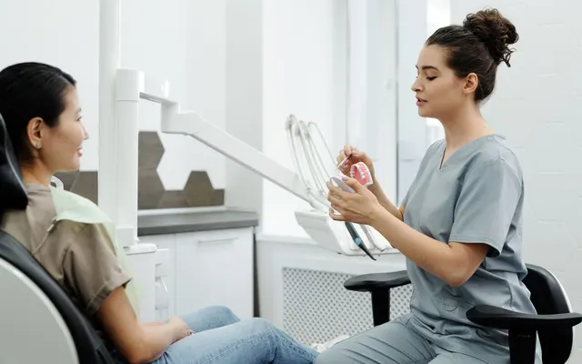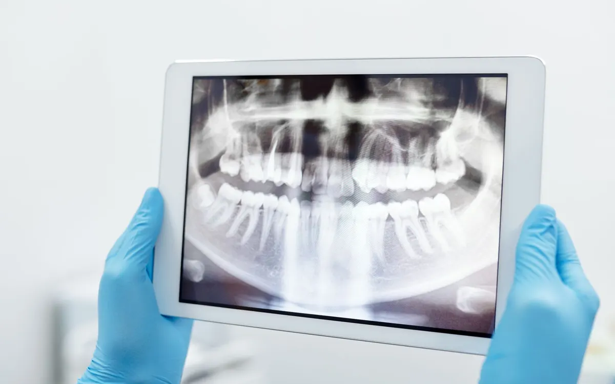
An image in the record is worth as much as what is written next to it: whose it is, which area, when, why it was taken and by whom. For a radiograph the why is written by the dentist before exposure, because the radioprotection rules do not allow unjustified exposures. For a photo the why is the purpose: care, teaching or promotion.
An image without a caption
The literature on dental records expects radiographs, study models and photographs to be labelled and dated, and the record to contain the clinical and radiographic findings together with the diagnosis and plan. The assistant may also record the radiographs taken, but responsibility for the record remains with the dentist (What's the deal with dental records, 2014 (opens in a new tab)).
The code of ethics (opens in a new tab) applies to the physician and to the dentist (art. 1). Art. 26 requires the objective data and the diagnostic and therapeutic activities carried out to be recorded in the record, with traceability of who wrote it. A radiograph is a diagnostic activity, and read literally the article covers it.
Decree 101/2020, article by article
The decree that transposed the European directive on ionising radiation has a title on medical exposures. These are the articles that touch on the record, as written in the text on Normattiva:
- Art. 157, justification. Diagnostic benefits must be weighed against potential harm, taking account of alternative techniques. The referring physician and the specialist use the diagnostic information already available, so as not to repeat unnecessary examinations.
- Art. 158, optimisation. Doses are kept as low as reasonably achievable, compatible with the diagnostic information needed. The choice of equipment and quality assurance programmes come into this.
- Art. 159, responsibility. Exposures are carried out on the motivated request of the referring physician. The text does not say in what form the request must be made.
- Art. 161, paragraphs 5 and 6, report. The report includes the information on the exposure. Pending the guidelines, this is the dose class, from I to IV, of the examination in question, identified on the basis of the type of examination and the indications of the medical physics expert.
- Art. 168, paragraph 1, recording. Investigations, treatments with radiation and the main technical parameters are recorded individually on electronic media, also for comparison with diagnostic reference levels.
Who, in the practice, is the "operator", the "installation manager" or the "medical specialist" within the meaning of the decree is established by the organisation of the practice, together with the lawyer and the medical physics expert. What matters here is what the record must be able to show, not the allocation of roles.
One line per examination
| Field | What to write | Why |
|---|---|---|
| Type | Intraoral, bitewing, panoramic, cephalometric, CBCT | The type changes the justification and the dose |
| Date | Day of exposure | Orders the history |
| Site | Teeth (FDI notation) or area | An examination without a site cannot be found |
| Justification | The clinical reason, written by you | It is what art. 157 requires you to be able to show |
| Parameters | kV, mA, time in seconds, DAP, as given by the machine | They say how it was carried out |
| Field of view | CBCT only, as given by the machine | The dose depends on it |
| Machine | Which machine | Parameters only make sense with the machine that produced them |
| Performed by | Who pressed the button | Traceability of who wrote it |
| Report | Yours, written by you | With the information on the exposure, art. 161 |
The question before exposure
The European guideline on paediatric dentistry requires an individual justification, specific to the patient (EAPD, 2020 (opens in a new tab)). For CBCT the 2023 Italian expert consensus says that the examination is justified when the limits of the two-dimensional one are clear (The Dedicated C.B.C.T., 2023 (opens in a new tab)), and a 2017 review adds that CBCT is used only when the question cannot be answered with conventional radiography at a lower dose (Role of CBCT, 2017 (opens in a new tab)). Writing the justification means writing that question: what you need to know, and why the examination chosen is the one with the least exposure that answers it.
The parameters
The Italian work on CBCT lists what governs dose and quality: current (mAs, that is mA times time), voltage (kV), voxel, field of view. The width of the field of view affects the dose and must be limited to the region of interest. DAP, dose area product, is used to compare the dose of different protocols. The register has fields for kV, mA, time, DAP and field of view. The voxel, if you want to keep it, goes in the report.
In the Fibonacci register
Each examination is a line, with the fields from the table, in the radiograph register. Without a justification it will not save. You copy the parameters from the machine, and the limits on the numbers only stop typing errors, a 700 instead of a 70. The register does not suggest the examination, does not calculate the dose, does not say whether it is high or low and does not derive the dose class for the report: you write the information on the exposure yourself.
Clinical photos
There are three uses for a dental photo: documentation, communication (with the patient, with the dental technician, with colleagues), promotion (Lazar and others, 2022 (opens in a new tab)). A 2020 survey puts it on a par with radiographs and models as a diagnostic tool, with written consent as best practice (Abouzeid and others, 2020 (opens in a new tab)). These are foreign studies: they describe practice, not an Italian obligation.
A clinical photo of the mouth, kept in the record, reveals health information and falls within data concerning health (definition in art. 4, point 15, of the GDPR (opens in a new tab), general prohibition on processing in art. 9). For why a photo taken for care does not require privacy consent, see health data in the dental practice.
In Fibonacci the photo is uploaded from the patient's file. On saving you indicate the area and, if needed, the treatment it refers to, and you declare the purpose: clinical, teaching or promotional. The location data of the shot is removed before saving, the image is encrypted, every opening goes into the access log. Arrows, circles and captions are saved separately and the original is never touched.
The shot can also declare the view, but the list provided is the one for the face (frontal, lateral, 45 degree oblique, dynamic), not the one for intraoral framing. The rule of repeating the same series at every visit still applies: two dates can only be compared if the shots are comparable. Consents for promotion are in health data in the dental practice; for how long to keep images there is retention of dental documentation.
A bitewing of 26 and 27
Data invented for the example, not of a real person.
| Field | Value |
|---|---|
| Patient | Test patient |
| Type, date | Bitewing, 12 March 2026 |
| Site | 26, 27 |
| Justification | Check for interproximal caries in an area with no visual access, last bitewing 18 months ago |
| Parameters | 70 kV, 7 mA, 0.16 s, DAP 40 mGy·cm² |
| Machine, performed by | Intraoral machine 1, practice assistant |
| Report | Radiolucent lesion distal of 26. Dose class: to be indicated with the guidance of the medical physics expert |
Before you file
- The site is in FDI notation
- The date is the date of exposure, not the date of upload
- The justification is written by you and states the clinical question
- The parameters are copied from the machine, not reconstructed
- For CBCT the field of view is there
- The report is yours and includes the information on the exposure
- Every photo has its purpose; for teaching or promotion there is a separate consent
- No image leaves the record via unprotected chat or email