For each tooth present you measure and record six sites: probing depth and recession in millimetres, bleeding on probing yes or no, plaque yes or no. Then, for each tooth, mobility and, on multi-rooted teeth, furcation. You do not record the clinical attachment level: it is the sum of probing depth and recession, and the software works it out itself. You choose the stage and grade of the periodontitis, because they do not emerge from the probing numbers.
The 2009 guideline: all patients, all teeth
The National guidelines for oral health in adulthood (opens in a new tab) (December 2009) state, in recommendation 3, that the diagnosis of periodontal disease is made by all dentists, on all patients and on all teeth. A detailed history, a local objective examination and a radiographic examination are required. Laboratory and microbiological tests are optional: the text justifies them when the result can change or refine the treatment. Strength of recommendation A, level of evidence I.
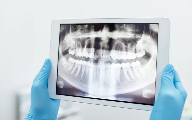
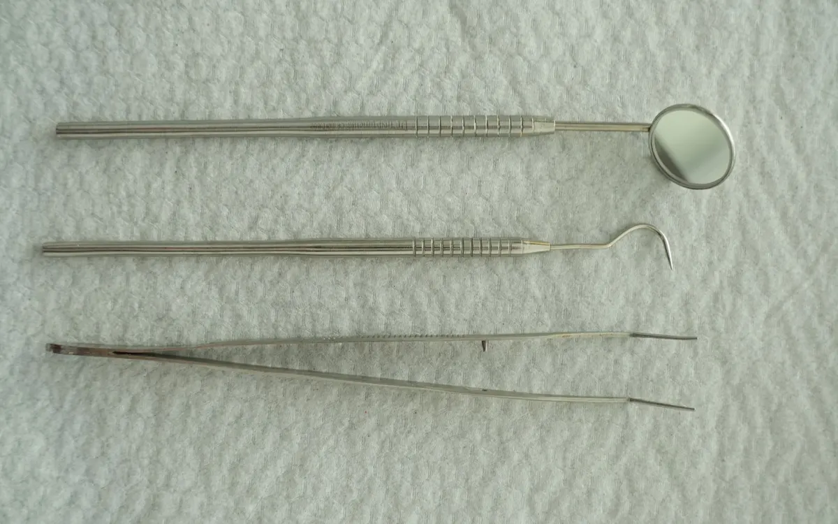
For the history, the text asks you to look for what influences onset and progression: smoking, drugs that increase gingival volume (nifedipine, diphenylhydantoin, ciclosporin), diabetes, certain rare systemic and genetic diseases. The objective examination looks at the gingiva and mucosae and, for the supporting tissue, records pocket depth, attachment level, furcation involvement, mobility. Then plaque and what retains it: calculus, caries, overhanging restorations, malpositions, crowding. Probing is done with a graduated probe on every surface of all teeth, and the periapical intraoral radiograph accompanies the measurement of pockets and attachment levels.
In the clinical record in Fibonacci, the dental history has fields for these facts, reported by the patient: drugs that increase gingival volume, diabetes and other systemic diseases, anticoagulants, heart valve prostheses.
Six sites per tooth, and what you record in each
Three sites on the buccal aspect (mesial, central, distal), three on the lingual or palatal aspect. This is the scheme used by research protocols: one registered in 2018 (opens in a new tab) lists mesio-buccal, buccal, disto-buccal and the three lingual ones.
| What | How it is defined | In Fibonacci |
|---|---|---|
| Probing depth (PPD) | distance between the free gingival margin and the bottom of the sulcus or pocket | you, in whole millimetres |
| Recession (REC) | distance between the cementoenamel junction and the gingival margin | you, in whole millimetres |
| Clinical attachment level (CAL) | sum of depth and recession | calculated, not recorded |
| Bleeding on probing | yes or no, site by site | you |
| Plaque | yes or no, site by site | you |
The definitions of depth, recession and clinical attachment come from a population study from 2021 (opens in a new tab). In the 2018 protocol, recession counts as zero when the cementoenamel junction is covered, and bleeding and plaque are a yes or a no.
The software calculates the clinical attachment level because a number derived from two others, typed a second time, can contradict them. The values are whole numbers and have limits, which stop typing errors and nothing else.
From the grid also come the percentage of sites that bleed and the percentage of sites with plaque, out of the total sites probed: these are the two numbers that studies report as percentages (the Dakar study from 2021 (opens in a new tab) gives them as group means).
A tooth marked absent in the odontogram drops out of the probing: the periodontal record uses the same grid, and nothing is recorded on a tooth that is not there.
Mobility and furcation, one per tooth
Mobility runs from 0 to 3. The four-grade scale is attributed to Miller; the 2026 study (opens in a new tab) that uses it describes it as grade 0 (physiological mobility), then slight, moderate, severe. Furcations also run from 0 to 3, with Hamp's classification (2023 protocol (opens in a new tab)). In Fibonacci, furcation is recorded only where there is more than one root, that is, on molars and upper first premolars. The software asks for the grade. How you measure it is up to you.
Why stage and grade remain with you
The 2017 World Workshop (opens in a new tab) consensus, published in 2018 by Europeans and Americans together, brought under «periodontitis» the forms previously called chronic and aggressive and described them with stages and grades. The stage depends above all on the severity at presentation and the complexity of management. The grade adds the biology: the rate of progression derived from the history, the risk of further progression, the possibility of unfavourable treatment outcomes, the risk that the disease or its treatment will weigh on general health.
There are two fields, stage from I to IV and grade from A to C, and you fill them in. The software does not deduce them, and the sources say why. In the table of the Dakar study, which applies the classification, the stage looks at the clinical attachment at the site of greatest loss, teeth lost to periodontitis and complexity factors (depth, furcations, mobility, bone defects, masticatory function). The grade looks at the ratio between bone loss and age, that between biofilm and destruction, and two modifiers, smoking and diabetes. Some of these data are in the radiographs, in the history, in the patient's record, not in the grid.
The same study recounts that, without a periodontal history and with non-computerised records, the direct criteria of progression (attachment or bone loss in the last five years) were difficult to use. This is why every save of the periodontal record is a new photograph and leaves the previous one intact: in two years, when you want to know how much attachment has been lost at a site, today's numbers must still be today's.
The 26 of 16 November (sample data, invented)
The clinical attachment level is calculated.
| Site | Probing (mm) | Recession (mm) | Attachment (mm) | Bleeding | Plaque |
|---|---|---|---|---|---|
| mesio-buccal | 3 | 0 | 3 | no | yes |
| buccal | 2 | 1 | 3 | no | no |
| disto-buccal | 4 | 1 | 5 | yes | yes |
| mesio-lingual | 3 | 0 | 3 | no | no |
| lingual | 2 | 0 | 2 | no | no |
| disto-lingual | 5 | 1 | 6 | yes | yes |
Mobility 0, furcation 1. Two sites out of six bleed, three out of six have plaque. Stage and grade cannot be derived from a single tooth: you record them when you also have radiographs and history.
To have in the record
- History: smoking, drugs that increase gingival volume, diabetes and other systemic diseases (these are the ones named by the Ministry guideline)
- Probing on all teeth present, six sites per tooth, with a graduated probe
- Depth and recession in millimetres, bleeding and plaque yes or no, for each site
- Mobility and furcation per tooth, where furcation makes sense
- Periapical intraoral radiograph linked to the probing: photos and radiographs in the dental record
- Stage and grade recorded by you, with the date, when you have all the data the classification requires
- Every follow-up is a new photograph, the old one is not corrected
- The treatment plan is in the estimate, item by item: treatment plan and dental estimate
How to keep documentation over time is in retention of dental documentation.